Evidence
Design, training and validation of Elavris Lite version 1.
Headline results
normal chest radiographs flagged as tuberculosis
radiographs with an annotated tuberculosis region flagged (98.5%)
tuberculosis-labeled radiographs without an annotated region flagged (63.8%)
Version 1 (epoch 8). Small internal validation sets. The held-out test round has not yet been run.
Why this design
Assessing tuberculosis on a chest radiograph requires two things at once: a judgement and a location. Elavris Lite produces both from a single multimodal model, so every positive finding arrives with an outline that can be inspected. Because the model accepts a text instruction alongside the image, the design leaves room to extend the same system to further findings as data allows.
How it works
-
Image encoding
A vision encoder converts the radiograph into visual features.
-
Language reasoning
A 7.4-billion-parameter language model reads those features together with a text instruction and replies in language. When it judges tuberculosis to be present, it emits a dedicated segmentation token.
-
Mask decoding
The representation at that token conditions a mask decoder, which produces a pixel-level outline of the suspected region. When no tuberculosis is suspected, no outline is produced.
Technical specification
| Architecture | Multimodal vision-language model with language-guided segmentation |
|---|---|
| Total parameters | 7.4 billion |
| Vision encoder | CLIP ViT-L/14 |
| Mask decoder | SAM-style decoder, image encoder frozen during fine-tuning |
| Outputs | Free-text reply and, when tuberculosis is suspected, a segmentation mask |
| Trainable parameters | About 204 million (roughly 2.8% of the model) |
| Fine-tuning | 8 epochs, 6,680 steps, about 10.5 hours on a single A100 GPU (40 GB), peak memory about 26 GB |
| Checkpoint selection | Rule fixed before any results were seen, validation data only; test split untouched. |
Training efficiency
Version 1 was fine-tuned from a pretrained multimodal base rather than trained from scratch. Fine-tuning updated about 204 million of 7.4 billion parameters and completed in roughly 10.5 hours on a single A100 GPU. This keeps each training round fast and inexpensive, which allows the model to be improved repeatedly as new data becomes available.
Training data
Version 1 was fine-tuned on chest radiographs from tuberculosis datasets, using a neutral instruction so that the image alone determines whether an outline is produced. Details of the training sources are not listed on this page.
Six examples, selected by a fixed rule
For each outcome, the first two validation images in filename order were taken. The rule was fixed before any results were generated. These examples illustrate behaviour and do not measure accuracy. Outputs are those of the model and have not been reviewed by a radiologist.
Normal radiographs, no finding reported
Normal validation radiograph 1

“No tuberculosis lesions are present in this chest radiograph.”
Normal validation radiograph 2

“This chest X-ray does not show findings consistent with tuberculosis. No segmentation is needed.”
Tuberculosis-labeled, flagged with a model outline
Tuberculosis-labeled radiograph 1 (flagged)
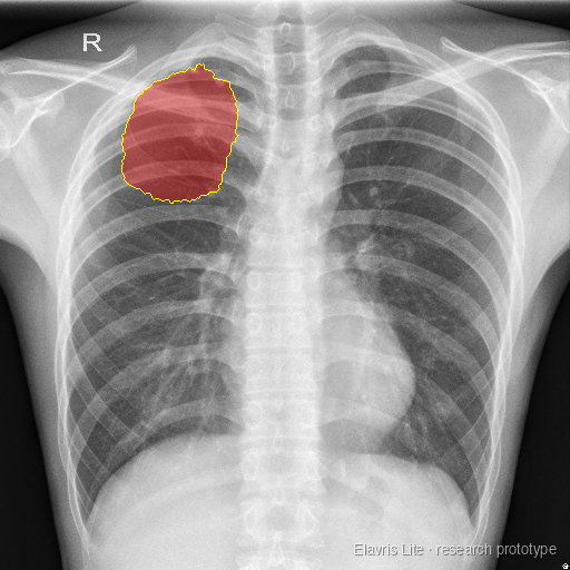
“Tuberculosis is present in this X-ray. The involved region is segmented.”
No reference outline exists for this image, so the highlighted region is model output only.
Tuberculosis-labeled radiograph 2 (flagged)
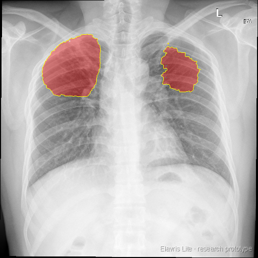
“I found tuberculosis involvement and outlined the affected regions.”
No reference outline exists for this image, so the highlighted region is model output only.
Tuberculosis-labeled, not flagged by version 1
Tuberculosis-labeled radiograph 3 (unflagged)

“This chest X-ray does not show findings consistent with tuberculosis. No segmentation is needed.”
Tuberculosis-labeled radiograph 4 (unflagged)

“This chest X-ray does not show findings consistent with tuberculosis. No segmentation is needed.”
Version 1 does not flag every tuberculosis case. These images define a target for the next round of data and training.
What we checked
| Check | Result | Interpretation |
|---|---|---|
| Normal radiographs flagged | 0 of 35 | No over-flagging of normal images in validation. |
| Radiographs with annotated tuberculosis region flagged | 197 of 200 | Flags tuberculosis consistently when the disease region is clearly annotated. |
| Tuberculosis-labeled radiographs without annotation flagged | 37 of 58 | The harder setting, and the focus of the next training round. |
All figures come from small internal validation sets. They are not proof of accuracy and are not compared with an earlier model.
Limitations and next steps
Data diversity. Version 1 was trained on small, mixed datasets.
Consistency across image sources. Sensitivity varied by source. On one source group, the model flagged 23 of 24 tuberculosis-labeled radiographs. On another, it flagged 13 of 33 while flagging none of the 35 normal radiographs, a conservative pattern. The sample is small, and the cause may be a source-specific shortcut, weaker labels or milder disease in those images.
Outline placement on non-square images. Outlines can sit slightly off on non-square images, about 0.08 lower in overlap score.
Held-out testing. The held-out test round has not yet been run.
Interested in evaluating Elavris Lite or contributing data?
Write to [CONTACT EMAIL] or see the roadmap to learn how version 1 develops.
Research prototype. Not a medical device. Not for diagnosis.